Confirmation
PR16: 12-Lead ECG Acquisition
Applicable To
Introduction
The 12-lead electrocardiogram is one of the most useful diagnostic tests in medicine and is a critical component in out-of-hospital care and decision-making. It allows paramedics to view the rhythm of the heart and provides important information about the state of blood flow to various regions of the heart.
Indications
- Suspicion of cardiac ischemia or rhythm disturbance
Contraindications
- As a diagnostic procedure, there are few absolute contraindications to 12-lead ECG acquisition; paramedics must ensure that the time needed to acquire a 12-lead ECG does not interfere with priority patient management tasks
Procedure
Procedure: Standard 12-Lead ECG
- Assemble required equipment. Connect electrodes to lead wires before placing them on the patient and connect the cables to the monitor. Ensure cables are not tangled.
- Prepare the patient’s skin as discussed below in 'Notes.'
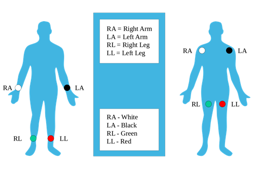
- Place the limb leads in the appropriate locations. RA and LA leads can be placed on the deltoids or wrists. RL and LL should be placed near the ankles (or alternatively, on the lower left leg). In all cases, ensure the leads are not positioned over bone.
- Landmark and place the precordial leads in their appropriate locations. Find the clavicle and identify the Angle of Louis as illustrated.
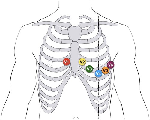
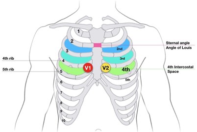
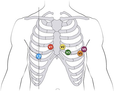
- V1 is located at the fourth intercostal space on the right of the sternum.
- V2 is also at the fourth intercostal space, but on the left side of the sternum.
- V3 is located between V2 and V4. For ease of placement, inexperienced operators should place V3 after V4 has been positioned.
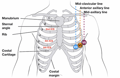
- V4 is placed at the fifth intercostal space on the mid-clavicular line. Generally, this will be inferior to the left nipple.
- V5 is also at the fifth intercostal space, but on the anterior axillary line.
- V6 is level with V5 on the mid-axillary line.
- Ask the patient to remain still, relax their body, not talk, and to breathe calmly. Press the “12 Lead” button on the LifePak 15. The monitor will prompt for an age and gender. Use the scroll wheel to enter the requested information and push the wheel to confirm each entry. (This information is critical for the machine interpretation algorithm and can also affect the processing of ECG signals by the LifePak 15. Paramedics must make every effort to enter this information accurately.)
- The monitor will attempt to acquire the ECG. If 'NOISY DATA – PRESS 12 LEAD TO ACCEPT' appears, attempt to identify the source of the problem (e.g., loose electrode contact, patient movement, tension on the lead wires, etc.) and correct the issue. The LifePak 15 will abandon the ECG recording if the noisy data persists for more than 30 seconds ('EXCESSIVE NOISE – 12 LEAD CANCELLED'); in this case, restart the acquisition process by pressing “12 Lead” again. If the noise persists, the LifePak 15 can be forced to acquire an ECG at the discretion of the ACP – press the “12 Lead” button when prompted to override.
- If the ECG is to be transmitted, press the “Options” button and select “Patient” from the menu. The patient’s name, PHN, or date of birth (in the “Patient ID” field), and the onset of pain (in the “Incident ID” field), can then be entered using the scroll wheel. The inclusion of this information is very important to minimize delays on arrival at hospital.
- To transmit the ECG, press the “Transmit” button. Select the desired ECG record and destination site, then select “Send” from the menu.
- ECGs may be re-printed by pressing “Options,” selecting “Print,” and then choosing the appropriate record.
Procedure: Posterior Leads
In some cases, a view of the posterior heart is needed, particularly in patients with marked precordial ST depression.
- Acquire a standard 12-lead ECG.
- Disconnect V4, V5, and V6 from their traditional placements.
- Using new electrodes, with the patient leaning forward:
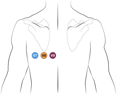
- Place the V4 electrode on the left posterior axillary line in the same plane as V6. This electrode becomes V7.
- Place the V5 electrode at the tip of the left scapula, in the same horizontal plane as V6. This electrode becomes V8.
- Place the V6 in the left paraspinal region, in the same plane as the other electrodes. This electrode becomes V9.
- Acquire the ECG.
- Once the LifePak 15 prints the ECG, mark V4, V5, and V6 with their new designations of V7, V8, and V9 on the rhythm strip.
Procedure: Right-Sided Leads
The right-sided chest lead is very helpful in diagnosing right ventricular infarctions.
- Acquire a standard 12-lead ECG.
- Disconnect V4 from its traditional placement.
- Using a new electrode, place V4 at the fifth intercostal space on the mid-clavicular line. This becomes V4R and is essentially the “mirror image” of V4 on the left chest.
- Acquire the ECG.
- Once the ECG is printed, mark V4 as V4R on the rhythm strip.
Procedure: Lewis Leads
The Lewis Lead ECG is used in order to have a specific and detailed view of atrial activity. This may be clinically useful when atrial flutter is suspected but not clearly demonstrated, or to detect P waves in a wide complex tachycardia.
- To create the Lewis Lead, move the right arm electrode to the 2nd intercostal space, right of the sternum. Move the left arm electrode to the 4th intercostal space, right of the sternum (traditionally the landmark for V1).
- Leave the lower limb leads in place.
- To read the Lewis Lead, print rhythm strip in 'Lead I'.
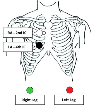
Image Credit: Life in the Fast Lane ECG Library
Notes
- 12-lead ECG acquisition is a relatively intimate procedure. Paramedics should strive to preserve patient dignity whenever possible by using gowns or towels.
- Tips for improved ECG quality:
- Skin preparation can significantly improve the quality of the ECG signal. Shave hair at the site of electrode placement whenever possible. An alcohol wipe can be used to help dry the skin when it is sweaty and a gauze pad can be used to rub the skin briskly to remove sweat, oil, and dead skin cells, improving contact.
- The conduction of ECG electrodes is improved as they warm. Consider ensuring that electrodes are stored at room temperature (up to body temperature is ideal).
- Do not press on the center of the electrode while applying it to the patient. Press around the circumference of the electrode to ensure proper adhesion.
- Patients should be supine or semi-recumbent during ECG acquisition. Their limbs should be fully supported.
- The Angle of Louis can be identified by placing a finger in the notch at the top of the sternum. Move the finger downward until a slight ridge or bump is felt, then slide the finger laterally to the patient’s right side to locate the second rib and the second intercostal space immediately below. Count down two more intercostal spaces; this is the fourth intercostal space and V1 is placed immediately adjacent to the sternum.
- V4 may be placed under the breast if necessary.
- In patients who have been resuscitated from cardiac arrest, wait at least ten minutes following sustained return of spontaneous circulation before attempting to record a 12-lead ECG.
Resources
References
- BCEHS STEMI Program Manual (link forthcoming)
- Life in the FastLane. ECG Lead Positioning Basics. [Link]